From microscopy to a digital model
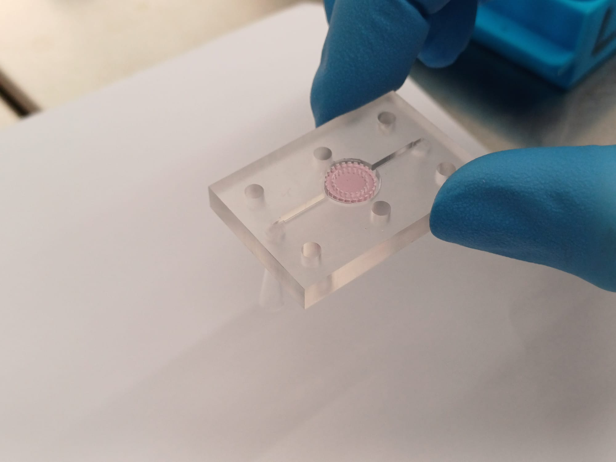
Brain Organ-on-Chip connects microscopy experiments with mathematical models of interactions between tumour cells and astrocytes. The objective is a reproducible pipeline that turns experimental observations into calibrated, predictive digital models on the geometry of the chip.
Methods
- Register microscopy images and regions of interest in the chip coordinate system.
- Detect and track cells in confocal image sequences; analyse displacement, speed and alignment.
- Reconstruct continuous cell-density fields from trajectories using kernel density estimation (KDE).
- Simulate cell densities, chemical signals and oxygen using partial differential equations and Feel++ HDG/CFPDE methods.
- Calibrate model parameters from comparable observables, with identifiability analysis and uncertainty quantification.
Collaboration
The work is part of an Italian collaboration in the D3 4Health context (Digital Driven Diagnostics, Prognostics and Therapeutics for Sustainable Healthcare), an Italian PNC/PNRR initiative on digital and biological twins for healthcare.
The presentation names G. Bretti, S. Bertoluzza, M. Pennacchio, A. De Giorgi and F. Gervaso, with CNR-NANOTEC among the collaborators. Christophe Prud’homme contributes for Cemosis / University of Strasbourg.
Development status
The July 2026 presentation reports available confocal data, an existing image-analysis and tracking workflow, and a brain organ-on-chip PDE case. Coordinate registration and micro-to-macro density reconstruction are under development. Three-dimensional reconstruction, observation-consistent calibration and Bayesian uncertainty analysis are further development objectives.